Anatomy Of Chest Muscles Female / Muscles Of The Chest And Upper Back / Muscles of the chest and their functions you have two mighty muscles on both sides of your chest:
Anatomy Of Chest Muscles Female / Muscles Of The Chest And Upper Back / Muscles of the chest and their functions you have two mighty muscles on both sides of your chest:. Pectoralis major muscle, associated fascia, and into the medial axillary wall. The serratus anterior starts from the inside of your shoulder blade, wraps around your side, and attaches onto the front of your ribcage. Here, we break down the anatomy of your chest muscles. Breast anatomy this image shows the anatomy of the female breast showing: Muscle anatomy chest 12 photos of the muscle anatomy chest anterior chest muscle anatomy, chest muscle anatomy and exercises, chest muscle anatomy male, chest wall muscle anatomy mri, female chest muscle anatomy diagram, human muscles, anterior chest muscle anatomy, chest muscle anatomy and exercises, chest muscle.
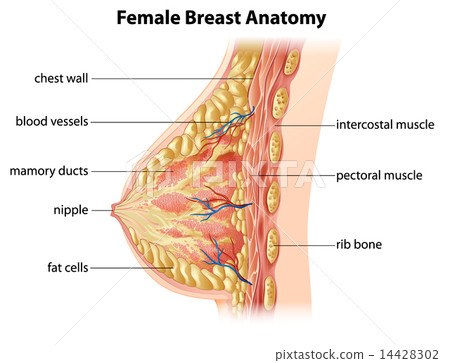
The diaphragm forms the upper surface of the abdomen. Thoracic wall the first step in understanding thorax anatomy is to find out its boundaries. The pectoral region is located on the anterior chest wall. Breast anatomy this image shows the anatomy of the female breast showing: Muscle anatomy chest 12 photos of the muscle anatomy chest anterior chest muscle anatomy, chest muscle anatomy and exercises, chest muscle anatomy male, chest wall muscle anatomy mri, female chest muscle anatomy diagram, human muscles, anterior chest muscle anatomy, chest muscle anatomy and exercises, chest muscle.
Female Breast Anatomy Stock Illustration 14428302 Pixta from en.pimg.jp No need to register, buy now! Thoracic wall the first step in understanding thorax anatomy is to find out its boundaries. Related posts of chest muscle in women body muscle anatomy practice exam. The pectoral region is located on the anterior chest wall. The diaphragm forms the upper surface of the abdomen. Chest muscle anatomy the pectoralis major muscles also known as the pecs are located on the front of the rib cage and form the major muscles of the chest. Find the perfect female chest anatomy stock photo. In humans, breast tissue begins to enlarge at puberty.
Each breast consists of tissue overlying the chest wall muscles (the pectoral muscles).
Posted on june 24, 2015 by admin. The abdomen (commonly called the belly) is the body space between the thorax (chest) and pelvis. Breast anatomy this image shows the anatomy of the female breast showing: Muscles the major muscle in the chest is the pectoralis major. The pectoralis major and the pectoralis minor, known collectively as your pecs. Pectoralis major muscle, associated fascia, and into the medial axillary wall. Here's how science can help you grow! See human chest anatomy stock video clips. Sternocleidomastoid muscle clavicle and ribs anatomy muscle anatomy chest sternocleidomastoid ribs anatomy chest muscles anatomy thorax rib muscles chest muscles chest anatomy illustration. The epidermis is the outermost layer that provides a protective waterproof seal over the body. 5 lymphatics of the breast syllabus p. The superior thoracic aperture found superiorly and the inferior thoracic aperture. Start with a pair of dumbbells extended above your chest.
Anyone who experiences chest pain or discomfort that lasts for several minutes or recurs should seek emergency. Muscles of the chest and their functions you have two mighty muscles on both sides of your chest: The serratus anterior starts from the inside of your shoulder blade, wraps around your side, and attaches onto the front of your ribcage. Chest a woman's chest — like the rest of her body — is covered with skin that has two layers. Muscles the major muscle in the chest is the pectoralis major.
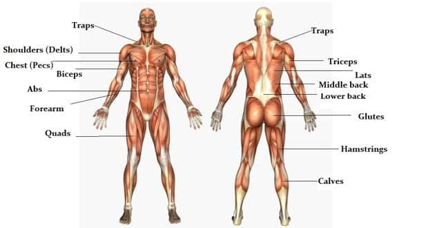
The Massive Muscle Anatomy And Body Building Guide You Always Wanted Thehealthsite Com from st1.thehealthsite.com Posted on june 24, 2015 by admin. In humans, breast tissue begins to enlarge at puberty. Chest muscle anatomy the pectoralis major muscles also known as the pecs are located on the front of the rib cage and form the major muscles of the chest. The pectoral region is located on the anterior chest wall. The image, which is of unknown origin, shows the female muscle system from the upper abdomen to the bottom of the neck. This diagram depicts picture of the female body 744×992 with parts and labels. The pectoralis major and the pectoralis minor, known collectively as your pecs. Fill out your shirt with a bigger, stronger, more powerful chest.
The major muscle in the chest is the pectoralis major.
The thorax has two major openings: Plus, how to target each to make them bigger and stronger. Fill out your shirt with a bigger, stronger, more powerful chest. The medical name for breast is mammary gland. It contains four muscles that exert a force on the upper limb: Sternocleidomastoid muscle clavicle and ribs anatomy muscle anatomy chest sternocleidomastoid ribs anatomy chest muscles anatomy thorax rib muscles chest muscles chest anatomy illustration. This diagram depicts female muscle anatomy chart. Huge collection, amazing choice, 100+ million high quality, affordable rf and rm images. Really lean bodybuilders have defined serratus muscles. The epidermis is the outermost layer that provides a protective waterproof seal over the body. Cross section anatomy of female chest and abdomen muscles stock illustration female muscle anatomy female muscular system full anatomical body diagram with muscle scheme vector illustration educational poster muscles in the chest anatomy female. Muscles the major muscle in the chest is the pectoralis major. Here, we break down the anatomy of your chest muscles.
The pectoral region is located on the anterior chest wall. The remaining part is made up of fatty tissue. Start with a pair of dumbbells extended above your chest. Sternocleidomastoid muscle clavicle and ribs anatomy muscle anatomy chest sternocleidomastoid ribs anatomy chest muscles anatomy thorax rib muscles chest muscles chest anatomy illustration. Related posts of chest muscle in women body muscle anatomy practice exam.
How To Avoid Breast Damage When Exercising An Interview With Professor Joanna Scurr from www.news-medical.net The pectoralis major, pectoralis minor, serratus anterior and subclavius. No need to register, buy now! The superior thoracic aperture found superiorly and the inferior thoracic aperture. Anyone who experiences chest pain or discomfort that lasts for several minutes or recurs should seek emergency. Female anatomy includes the external genitals, or the vulva, and the internal reproductive organs. Thoracic wall the first step in understanding thorax anatomy is to find out its boundaries. Find the perfect female chest anatomy stock photo. The abdomen (commonly called the belly) is the body space between the thorax (chest) and pelvis.
Thoracic wall the first step in understanding thorax anatomy is to find out its boundaries.
Start with a pair of dumbbells extended above your chest. This diagram depicts female muscle anatomy chart. The epidermis is the outermost layer that provides a protective waterproof seal over the body. It contains four muscles that exert a force on the upper limb: The abdomen (commonly called the belly) is the body space between the thorax (chest) and pelvis. Female muscle groups anatomical fitness vector illustration, sports muscle diagram, most important muscles of an athletic black man, anterior and posterior view pectoralis major muscle, muscles of chest, beautiful design of human anatomy front side with. Fabian identifying the muscles and landmarks of the abdomen and chest. Three dimensional view of the female reproductive system, full frontal view. Sternocleidomastoid muscle clavicle and ribs anatomy muscle anatomy chest sternocleidomastoid ribs anatomy chest muscles anatomy thorax rib muscles chest muscles chest anatomy illustration. The medical name for breast is mammary gland. The image, which is of unknown origin, shows the female muscle system from the upper abdomen to the bottom of the neck. The remaining part is made up of fatty tissue. The major muscle in the chest is the pectoralis major.


0 Komentar